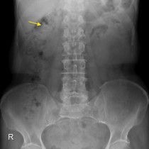
During the first stage, on the normally positioned left renal unit a ureteral stent was placed initially. The patient underwent staged bilateral RIRS under general anesthesia. The left kidney was positioned normally in the renal fossa. Elongated right ureter of TK showed unobstructed drainage in excretory phase of the CT urogram. The TK was malrotated, with the renal hilum facing anteromedially (Fig. Mild caliectasis of the upper pole calyces with normal sized pelvis in the right TK was noted (Fig. A well-defined soft tissue density structure measuring 23 × 18 mm was noted lateral to and abutting right psoas muscle at L4 level (suggestive of intraabdominal testis in the given case scenario) (Fig. The right kidney was displaced superiorly with simultaneous upward displacement of right lobe of liver and colon (Fig. CT also showed smooth elevation of diaphragm on right side and was suggestive of diaphragmatic eventration. 1a and b), and left partial staghorn calculus was measuring 24 × 18 mm (Hounsfield unit 1135) (Fig. Right partial staghorn calculus was measuring 36 × 20 mm (Hounsfield unit 1340) (Fig. The right testis was not palpable in the scrotum, whereas his left testis was palpable in the scrotum and was normal in volume.ĬT (Computed tomography) abdomen showed bilateral partial staghorn calculi. There was no history of any surgery or significant trauma in the past. There was no history of respiratory tract infections in the past. There were no complaints of vomiting, difficulty in breathing or chest pain.

A 23-year-old male presented with the complaints of back pain of three months of duration.


 0 kommentar(er)
